Careful planning and a detailed understanding of oral anatomy are crucial for successful dental implant placement. Diagnostic imaging is essential for this, offering a detailed view of the jawbone and surrounding structures. Proper imaging ensures a safer procedure, better outcomes, and a longer-lasting smile.
Cone Beam Computed Tomography (CBCT)
Cone Beam Computed Tomography, or CBCT, has become the gold standard for dental implant planning. Unlike traditional two-dimensional X-rays, a CBCT scanner produces a three-dimensional image of the teeth, soft tissues, nerve pathways, and bone in a single scan. This comprehensive view is invaluable for complex procedures like implant placement.
How CBCT Works
During a CBCT scan, a cone-shaped X-ray beam rotates around the patient’s head. In less than a minute, the machine captures hundreds of images, or “views,” from different angles. Sophisticated software then reconstructs these images into a single 3D model. This model can be manipulated on a computer, allowing the dental professional to view the jaw from any angle and take precise measurements.
Advantages of CBCT for Implant Planning
The level of detail provided by CBCT scans offers several significant benefits for implant placement:
- Precise Measurements: CBCT allows for highly accurate measurements of bone height, width, and density. This is crucial for selecting the correct size and type of implant to ensure a stable foundation.
- Identification of Vital Structures: The 3D images clearly show the location of important anatomical structures, such as the inferior alveolar nerve in the lower jaw and the maxillary sinus in the upper jaw. Knowing the exact position of these structures helps prevent potential complications like nerve damage or sinus perforation during surgery.
- Virtual Implant Placement: With specialized software, dentists can perform a “virtual surgery” on the 3D model. They can place a digital version of the implant into the bone to test its fit and position before the actual procedure. This level of pre-planning leads to more predictable and successful outcomes.
- Assessment of Bone Quality: CBCT provides information about the quality of the bone, not just the quantity. This helps determine if a bone graft is needed to support the implant.
Limitations of CBCT
While CBCT is a powerful tool, it does have some limitations. The radiation dose, while significantly lower than medical CT scans, is higher than that of standard dental X-rays. Therefore, its use is typically reserved for cases where the benefits of detailed 3D imaging outweigh the risks. Additionally, the cost of a CBCT scan is higher than for other dental imaging methods.
Panoramic Radiography
Before the widespread adoption of CBCT, panoramic radiography was the primary imaging tool for initial implant assessment. A panoramic X-ray provides a flat, two-dimensional overview of the entire mouth, including both the upper and lower jaws, teeth, and temporomandibular joints (TMJs).
How Panoramic Radiography Works
To capture a panoramic image, the X-ray machine rotates around the patient’s head. The patient remains still while the machine creates a single, wide-view image. This process is quick, simple, and exposes the patient to a relatively low dose of radiation.
Role in Implant Planning
Panoramic radiographs are excellent for a broad initial assessment. They help the dentist:
- Evaluate Overall Oral Health: A panoramic image can quickly identify issues like widespread bone loss, existing infections, or unerupted teeth that may need to be addressed before implant surgery.
- Screen for Potential Issues: It provides a good general overview of the available bone height and the proximity of major anatomical structures, like the maxillary sinuses.
- Initial Consultation Tool: Due to its convenience and low cost, it is often the first image taken during a consultation to determine if a patient is a potential candidate for dental implants.
Limitations of Panoramic Radiography
The main drawback of panoramic radiography is its two-dimensional nature. This leads to several limitations:
- Lack of Depth Information: A panoramic X-ray cannot show the width or thickness of the jawbone. This is a critical piece of information needed to select an appropriately sized implant.
- Geometric Distortion: The imaging process can cause magnification and distortion, leading to inaccurate measurements of bone height.
- Superimposition of Structures: Because it’s a 2D image, anatomical structures can overlap, making it difficult to clearly see the exact position of nerves or the sinus floor.
Due to these limitations, a panoramic X-ray alone is generally not sufficient for the final planning of an implant case. It is often used as a preliminary screening tool, followed by a more detailed 3D scan like a CBCT if the patient is a good candidate.
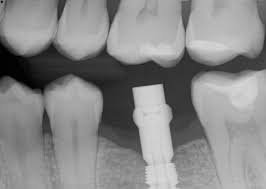
Periapical Radiographs
A periapical radiograph is a small, detailed X-ray that focuses on a few specific teeth. It captures the entire tooth, from the crown to the tip of the root, as well as the surrounding bone.
How Periapical Radiographs Work
To take a periapical X-ray, a small film or digital sensor is placed inside the mouth, and the X-ray machine is positioned outside. It is a quick and straightforward procedure that provides a high-resolution image of a localized area.
Role in Implant Planning
While not used for the initial broad assessment, periapical radiographs are very useful for specific tasks in the implant process:
- Detailed View of a Specific Site: If there is a question about the health of the bone or adjacent teeth in a specific implant location, a periapical X-ray offers the highest level of detail.
- Post-Operative Evaluation: After an implant has been placed, a periapical radiograph is often used to check its position and ensure it is integrating properly with the bone (a process called osseointegration).
- Long-Term Monitoring: During regular check-ups, these small X-rays are perfect for monitoring the health of the bone around the implant over many years.
Limitations of Periapical Radiographs
The primary limitation of periapical X-rays is their small field of view. They cannot provide an overview of the entire jaw or give information about the width of the bone, making them unsuitable as the sole imaging tool for implant planning.
Comparing the Imaging Methods
Each diagnostic imaging method has a distinct role in the dental implant journey. Highly skilled prosthodontists like those in Las Vegas often use a combination of these techniques to ensure the best possible result.
| Feature | CBCT | Panoramic Radiograph | Periapical Radiograph |
| Dimensionality | 3D | 2D | 2D |
| Primary Use | Detailed pre-surgical planning | Initial screening and overall assessment | Detailed local view and post-op monitoring |
| Key Advantage | Accurate measurements, 3D visualization | Broad overview of the entire mouth | High resolution of a small area |
| Limitation | Higher radiation dose and cost | Lack of depth information, potential distortion | Limited field of view |
| Bone Width Info | Yes | No | No |
| Nerve/Sinus View | Precise 3D location | General 2D approximation | Limited to the specific area captured |
A common workflow might involve taking a panoramic X-ray at the first consultation for a general overview. If the patient seems to be a good candidate, a CBCT scan would then be ordered for detailed, site-specific planning. Periapical X-rays would be used later to monitor the implant’s health.
Conclusion
Meticulous planning is key to successful dental implants. Diagnostic imaging, including CBCT for detailed pre-surgical planning and panoramic/periapical radiographs for initial assessment and long-term follow-up, provides essential information for safe, accurate, and predictable implant placement.