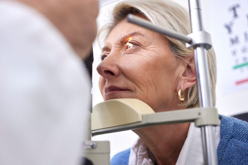
Diabetic retinopathy is one of the most common and serious complications of diabetes, affecting the eyes and potentially leading to vision loss if left untreated. It occurs when high blood sugar levels damage the small blood vessels in the retina — the light-sensitive tissue at the back of the eye responsible for clear vision.
With diabetes rates rising globally, understanding diabetic retinopathy has never been more important. Early detection, regular eye exams, and proper management of blood sugar can prevent severe damage and preserve vision.
What Is Diabetic Retinopathy?
Diabetic retinopathy (DR) is a progressive eye disease caused by long-term high blood sugar (glucose) levels that weaken and damage retinal blood vessels. Over time, these vessels can swell, leak, or close off completely, reducing blood flow to the retina. In some cases, abnormal new blood vessels grow — a process called neovascularization — which can cause scarring and vision problems.
The condition typically affects both eyes and develops gradually, often without noticeable symptoms in its early stages. However, as it advances, it can lead to blurred vision, floaters, or even blindness if not managed properly.
Types of Diabetic Retinopathy
There are two main stages of diabetic retinopathy:
1. Non-Proliferative Diabetic Retinopathy (NPDR)
This is the early stage of the disease. The walls of tiny blood vessels in the retina weaken and develop small bulges called microaneurysms, which may leak fluid and blood into the retina.
Symptoms are often mild or absent at this stage, but the retina may begin to swell, especially in the macula (the central part of the retina responsible for sharp vision), leading to diabetic macular edema (DME).
Key Features of NPDR:
- Microaneurysms (tiny bulges in blood vessels)
- Retinal swelling (edema)
- Small hemorrhages or leaking fluid
- Hard exudates (yellow deposits of fat or protein)
As NPDR progresses, blood vessels may become blocked, reducing oxygen supply to the retina.
2. Proliferative Diabetic Retinopathy (PDR)
This is the advanced stage of the disease. When retinal areas become oxygen-deprived, the body tries to compensate by growing new, abnormal blood vessels. However, these vessels are fragile and prone to bleeding into the vitreous (the gel-like substance inside the eye).
If left untreated, PDR can lead to:
- Vitreous hemorrhage – causing sudden vision loss.
- Retinal detachment – scar tissue pulls the retina away from the back of the eye.
- Neovascular glaucoma – new blood vessels block fluid drainage, increasing eye pressure.
PDR poses a serious threat to vision and requires immediate medical intervention.
Causes and Risk Factors
Diabetic retinopathy is primarily caused by prolonged high blood sugar levels, which damage the tiny blood vessels that nourish the retina. Over time, these vessels become blocked or leaky, disrupting normal vision.
Common Risk Factors Include:
- Duration of Diabetes: The longer you’ve had diabetes, the higher your risk.
- Poor Blood Sugar Control: Consistently high glucose levels accelerate retinal damage.
- High Blood Pressure (Hypertension): Increases the risk of vascular damage.
- High Cholesterol: Can worsen leakage and swelling in the retina.
- Pregnancy: Hormonal changes may increase risk in diabetic women.
- Kidney Disease: Often linked with worsening diabetic eye complications.
- Smoking: Damages blood vessels and accelerates disease progression.
Symptoms of Diabetic Retinopathy
In its early stages, diabetic retinopathy may not cause any symptoms. As the disease advances, however, vision problems become more noticeable.
Common Symptoms Include:
- Blurred or fluctuating vision
- Dark or empty areas in the visual field
- Floaters (dark spots or strings that move across vision)
- Difficulty seeing at night
- Colors appearing faded or washed out
- Sudden vision loss (in severe cases due to bleeding or retinal detachment)
If you have diabetes, it’s vital to have regular eye exams, even if you notice no changes in vision. Early detection allows for timely treatment and a much better prognosis.
Diagnosis of Diabetic Retinopathy
Diagnosing diabetic retinopathy involves a comprehensive dilated eye exam conducted by an optometrist or ophthalmologist.
Diagnostic Tests May Include:
- Visual Acuity Test: Measures clarity of vision.
- Dilated Eye Exam: Allows doctors to examine the retina and optic nerve for abnormalities.
- Optical Coherence Tomography (OCT): Provides detailed cross-sectional images of the retina to detect fluid accumulation or swelling.
- Fluorescein Angiography: A special dye is injected into a vein, highlighting leaking or blocked blood vessels under a specialized camera.
- OCT Angiography (OCTA): A noninvasive imaging method that maps blood flow in the retina.
These tests help determine the severity and type of diabetic retinopathy, guiding treatment decisions.
Treatment for Diabetic Retinopathy
The goal of treatment is to slow disease progression, prevent vision loss, and, when possible, restore sight. The appropriate approach depends on the disease stage and the presence of macular edema.
1. Blood Sugar and Lifestyle Management
In early stages (mild NPDR), controlling diabetes may be sufficient to prevent worsening. Key steps include:
- Maintaining healthy blood sugar levels.
- Managing blood pressure and cholesterol.
- Following a balanced diet and exercising regularly.
- Avoiding smoking and alcohol.
Regular monitoring and adherence to medication prescribed by your physician are essential.
2. Anti-VEGF Injections
In advanced cases or when macular edema is present, anti-VEGF therapy is one of the most effective treatments.
Anti-VEGF medications, such as Ranibizumab (Lucentis), Aflibercept (Eylea), or Bevacizumab (Avastin), block the effects of vascular endothelial growth factor (VEGF), a protein responsible for abnormal blood vessel growth and leakage.
Benefits:
- Reduces macular swelling.
- Improves or stabilizes vision.
- Slows disease progression.
Patients may need repeated injections every few weeks or months, depending on the severity and response.
3. Focal or Grid Laser Photocoagulation
Laser therapy is used to seal leaking blood vessels and prevent further fluid accumulation. It’s especially effective in treating diabetic macular edema.
- Focal Laser: Targets specific leaking vessels.
- Grid Laser: Treats diffuse areas of swelling in the macula.
Although it may not fully restore lost vision, it helps stabilize eyesight and prevent further deterioration.
4. Panretinal Photocoagulation (PRP)
In proliferative diabetic retinopathy, panretinal photocoagulation is used to shrink abnormal new blood vessels.
A laser is applied across the peripheral retina to reduce oxygen demand, thereby stopping the growth of fragile new vessels.
Advantages:
- Prevents severe bleeding or retinal detachment.
- Reduces the risk of blindness.
Some patients may experience mild peripheral vision loss or night vision difficulty as side effects.
5. Vitrectomy Surgery
For advanced PDR cases with vitreous hemorrhage or retinal detachment, a surgical procedure called vitrectomy may be required.
Procedure Overview:
- The vitreous gel filled with blood or scar tissue is removed.
- The space is replaced with a clear saline solution or gas bubble.
- The retina is reattached if necessary.
Vitrectomy can restore and preserve vision when other treatments are not effective.
Preventing Diabetic Retinopathy
While treatment options are effective, prevention remains the best approach. You can lower your risk significantly by managing your overall health.
Prevention Tips:
- Control Blood Sugar: Keep glucose within target range through diet, medication, and monitoring.
- Manage Blood Pressure and Cholesterol: Follow your doctor’s advice for a heart-healthy lifestyle.
- Regular Eye Exams: Schedule a dilated eye exam at least once a year — or more frequently if advised.
- Quit Smoking: Smoking increases the risk of diabetic complications, including eye disease.
- Exercise Regularly: Physical activity improves circulation and helps manage blood sugar.
Early intervention can stop or slow diabetic retinopathy before it threatens your vision.
Outlook and Living with Diabetic Retinopathy
Thanks to advances in medical technology and early screening, blindness from diabetic retinopathy is largely preventable today. With regular eye care, diabetes management, and timely treatment, most patients maintain good vision throughout their lives.
If you notice any changes in your vision, even subtle ones, consult an eye care professional immediately. Prompt action can make the difference between preserved sight and permanent vision loss.
Conclusion
Diabetic retinopathy is a serious but manageable eye condition that reflects the broader effects of uncontrolled diabetes. By maintaining healthy blood sugar levels, scheduling routine eye exams, and seeking timely treatment, individuals with diabetes can protect their vision for years to come.
Modern treatments such as anti-VEGF injections, laser therapy, and vitrectomy have revolutionized diabetic eye care — transforming a once-devastating complication into a controllable condition.
Your eyes are irreplaceable. Prioritize your vision, manage your diabetes, and take proactive steps today to ensure a clearer, brighter tomorrow.


